Review of PEEK Material & Silicon Nitride Ceramic Spinal Implants in the Same Patient
SUMMARY
A 66 year old female patient underwent two separate spinal surgeries, the first using two PEEK (polyetheretherketone) spinal implants and the second, three years later, using a Silicon Nitride ceramic (Si3N4) implant. This review compares the two materials post-operatively.
PEEK MATERIAL – DIAGNOSIS & FIRST SPINAL SURGERY
Patient had a 30 year history of chronic back pain. She reported two ruptured discs, which had been treated conservatively after an episode that left her unable to walk and on bedrest for two months. For five weeks she had experienced bilateral pain in her lower back, which radiated to both hips and down the posterior quadriceps, being most intense in her buttocks. There was tingling across her lower back, legs gave out intermittently, left leg pain was greater, and she could not stand for longer than 30-40 minutes. No bowel or bladder incontinence.
Patient had Von Willebrand Disease, hypercholesterolemia, hypertension, and diabetes mellitus that was under diet control. She was a social drinker and smoked 1 ½ packs per day. Radiologic examination revealed; severe facet disease bilateral at L5-S1, moderate facet disease at L4-L5 with left foraminal narrowing, and mild facet disease at L3-L4.
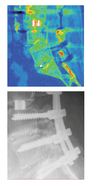
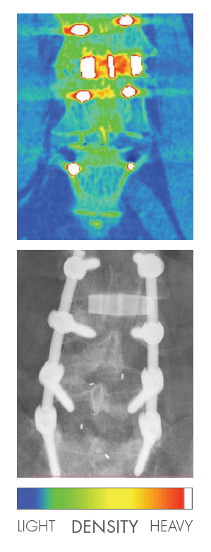
After a course of therapy and treatment with little marked improvement, surgical intervention was performed with a laminotomy and decompression of the L4, L5, and S1 nerve roots and a discectomy of L4-L5-S1. Ten millimeter PEEK material implants were placed obliquely across the midline with autologous posterior element bone packed in and around the spacer.
SILICON NITRIDE CERAMIC – DIAGNOSIS & SECOND SPINAL SURGERY
Three years later, the patient returned with intractable back pain which had lasted for 8 weeks. Radiographs indicated no evidence of bony incorporation of the PEEK devices. A broad based protruding herniation of the L3-L4 intervertebral disc existed, with moderate spinal stenosis and narrowing of the right intervertebral neural foramen, causing compression of the right L3 nerve root as it exited.
Additionally, there was compression of the left L4 nerve root as it passed through the lateral recess, a result of the bulging disc, osteophytes and left facet arthropathy.
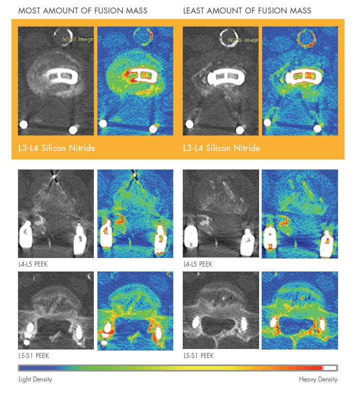
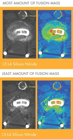
After conservative treatment a laminectomy of L3-L4 was performed, along with a discectomy from the right side where the patient experienced pain. An 11mm silicon nitride ceramic TL device was positioned midline. The Valeo™ TL device and space were packed with autologous posterior element bone graft.
CONCLUSION
Comparing PEEK to Silicon Nitride in these 7.4 year (PEEK material) and 4.4 year (silicon nitride ceramic) postoperative radiographs – it is apparent that silicon nitride fosters superior bone growth and bone density, despite having been implanted three years later.
Given the patient’s comorbidities and these results, silicon nitride should provide a clinician with significant outcome advantages over PEEK material.
For More Information Please Reach Out on the Contact Form.